


25
April
Warroad Wellness
Thursday


Imaging services are available to provide a variety of images of the human body for prevention, diagnosis, and treatment of disease. For questions regarding Imaging/Radiology services contact 218-463-4753.
Computed-tomography (CT) scan is a special type of x-ray that provides a more detailed view of organs, blood vessels, bones and soft tissue than regular x-rays. CT scans take multiple images from different angles. The result is a series of images that look like “slices”, which can then be reconstructed on a computer to provide a three-dimensional view of the area that was scanned. These views help your doctor to better diagnose problems such as cancer, heart disease, back problems, blood clots and other disorders.
LifeCare Medical Center’s CT scanner is a 128 system that has many built-in features to enhance our patient’s experience. The system uses the lowest possible radiation dose to provide the most detailed scans. Radiation dose is monitored via a system that alerts the technologist if a standard is exceeds. In addition, the system is much faster, allowing us to capture images from a larger area in a shorter amount of time; usually only requiring a 7 second breath hold. Finally, in cases where contrast is needed, our new system relies on weight-based dosing. All of these enhancements allow us to further personalize your healthcare experience while still providing top-notch scans to our providers.
Preparing for a CT – Your healthcare provider will give you a list of instructions to follow for your particular exam type. If you are having a scan of the abdominal or pelvic area, you might be asked to drink a contrast beverage the night before and 1 to 2 hours prior to the exam. The oral contrast has a vanilla flavor, and most patients do not find it unpleasant.
You may have to arrive before your scan to have blood drawn to check the functioning of your kidneys. Well functioning kidneys are necessary to help filter and excrete the IV contrast that may be used in your exam.
Clothing- Depending on the type of scan you are having, you may be asked to change into a gown. In some cases, you may be able to wear your regular clothing, so we encourage you to wear loose-fitting, comfortable clothes that have no metal.
Scanning – A technologist will position you on the examination table. Once in position, you will be moved through a doughnut shaped ring, called a gantry, which is the scanner. The technologist will have you in full view at all times and be in constant communication via a two-way microphone. Patients can expect the entire CT process to take from 30 to 90 minutes.
During your exam, you may be given contrast through an IV to highlight blood vessels. When the IV is administered, you may feel warm, have facial flushing or have a metallic taste in your mouth as it is being injected. These sensations generally last less than a minute and are normal.
Occasionally, patients may experience an allergic reaction to the contrast. Let the technologist know in advance if you have any known allergies to iodine, or if you experience any itching, rash, hives or difficulties with breathing or swallowing during the scan. Also inform the technologist if you have kidney problems, any diabetes medications you are taking, and if you are pregnant or think you may be before any contrast is administered.
After the Scan – You can return to your normal activities and routine diet right away. Any contrast you were given will pass naturally through your body. If you drank oral contrast, you may notice a milky color to your bowel movements, which is a normal occurrence. You should also drink more fluids after the scan to help your body “flush” the IV contrast out.
Images are made available for radiologist and provider review immediately following the exam in our PACS system. Results of routine exams are provided to the ordering provider by the end of the next business day. Emergent exams typically have results available within 30 minutes of the images being available. All images can be burned to CD upon patient request for easy transport to outside providers, or sent to networked facilities online.
A bone density test measures the density of your bones to estimate their strength. Used mostly in screening of post-menopausal women but can be used in younger women and men to detect bone loss.
Your examination will be taken in either an upright manner or with you lying down. Some examinations require the technologist to take several images and you may be repositioned for additional views.
For fluoroscopy exams, you will be instructed to not eat or drink the night before. We generally schedule exams such as UGI, barium enemas and esophagus studies in the morning for this reason. During the exam, the radiologist will use “live” x-ray to view your anatomy, and then save the images. You may be asked to drink an oral contrast beverage, depending upon the exam. You will be given complete instructions for your exam at the time of scheduling.
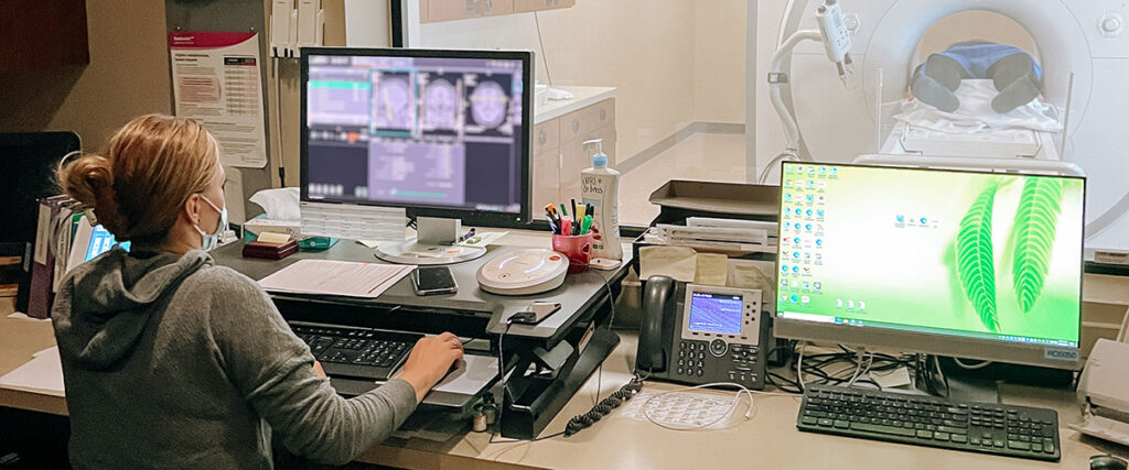
Magnetic resonance imaging (MRI) is a type of scan that uses a large, powerful magnet, radio waves and a computer to provide detailed images of various internal structures, such as bones, organs, blood vessels and other soft tissues. The MRI scanner looks like a large tube that is open on both ends with an examination table that moves into the opening.
LifeCare’s MRI has a larger opening. This helps to accommodate claustrophobic patients. Every effort is made by staff to make our patients as comfortable as possible during their exam. Music of your choice will be delivered to headphones during your scan. If you require special assistance for your exam, please feel free to call us at 218-463-4753 so special arrangements can be made.
Preparing for an MRI –
Prior to your MRI, your technologist will assist you in completing the MRI Screening Form. It is a good idea to review this form ahead of time to make sure you do not have factors that could make you ineligible for the test. Your healthcare provider will give you a list of instructions to follow for your particular exam type. Contact your healthcare provider or the LifeCare Imaging Department if you have any questions.
Clothing and Other Accessories – You will be asked to change into a gown. In some cases, you may be able to wear your regular clothing, so you’re encouraged to wear loose-fitting, comfortable clothing that do not have any metal fastenings, such as snaps, zippers or hooks.
You cannot enter the MRI unit with any metal or electronic items. Some of these items include:
You will be asked to complete a safety screening form and answer questions pertaining to your medical history to make sure it is safe for you to enter the MRI suite.
Contrast (Dye) – Some scans require the use of a contrast agent. The contrast or “dye” used is called gadolinium and it makes the internal structures of the body more visible on a scan. If your scan requires contrast, the technologist will start an IV to inject the gadolinium. Patients older than 60 years of age will need lab work drawn to see how well their kidneys are functioning before receiving the dye.
Scanning – The technologist will help to position you as comfortably as possible on the examination table. A device called an imaging coil will be placed over or under you. The table will then move into the magnet. From the control area, the technologist will stay in constant contact with you, both visually and through a two way microphone. During the scan, you will hear rapidly repeating, loud thumping noises coming from the scanner. To protect your ears, head phones with music and ear plugs will be provided. The scan will last from 20 minutes to an hour, depending on the area to be imaged.
Claustrophobia – Some people with claustrophobia have difficulties with MRI scans. We use a variety of methods to help combat these anxieties including music of your choice, special cushioning, and alternative positioning. If you are very apprehensive, please discuss your concerns with your doctor before your examination. Your doctor can prescribe a mild sedative or discuss other possible alternatives with you.
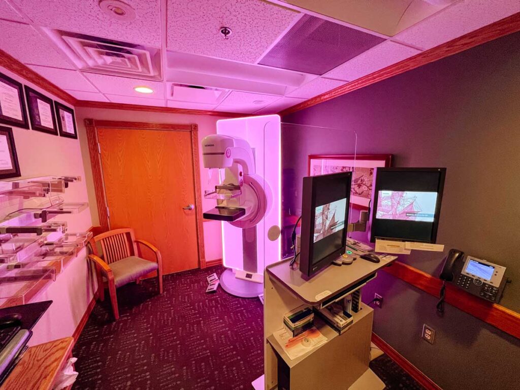
At LifeCare, we offer advanced digital Tomosynthesis (3D) mammography. Mammography provides an x-ray examination of breast tissues. It uses a very low dose of radiation and is considered very safe. Digital Tomosynthesis mammography creates three-dimensional images of the breasts and offers exceptional image clarity and detail by imaging the breast in several “layers.” The results include fewer biopsies and additional callbacks as well as increased accuracy.
The goal of screening mammography is to detect cancer when it is still too small to be felt by a doctor or the patient. Early detection of small breast cancers by screening mammography greatly improves a woman’s chances for successful treatment.
The American College of Radiology currently recommend that a woman of average risk have a mammogram every year starting at age 40. If you have a family history of breast cancer, your doctor may recommend beginning screening mammograms earlier. If you are already experiencing symptoms or notices changes such as nipple discharge or a lump in the breast, a mammogram should be performed.
Need help paying for a mammogram?
Through a grant provided by SAGE screening has funds available to help pay for the cost of a digital mammogram. Applying for the funds is simple because the ultimate goal is to ensure every woman who needs a mammogram gets one as early as possible.
For information about obtaining a free or reduced cost mammogram, contact LifeCare Imaging at 218-463-4753.
Preparing for a mammogram – You will be asked to undress above the waist and will be given a cape to cover you, wear clothing that will be easy to change out of. Do not wear deodorant or talcum powder on the day of the exam. The ingredients in these products can cause artifacts in the images. If you’ve had mammograms taken at other facilities, bring those films or records with you, or request to have them sent from the other facilities. These provide the radiologist with something to compare the current images.
What to expect during a mammogram
The technologist will help position the breast on the platform of the mammography machine. A plastic plate will be lowered to compress and flatten the breast tissue. This helps to separate the breast tissue and hold it immobile while the image is created.
The entire procedure only takes about 10 minutes.
Sometimes the radiologist will require additional images of the breast to better evaluate a specific area that may have changed since a previous exam or is suspicious in appearance. The radiologist may do a focal compression mammogram or they may recommend a breast ultrasound or a stereotactic biopsy.
Results
A letter indicating the results of your mammogram will be mailed to you within a week of your exam, and the results will be sent to your physician. You and your doctor will be notified by phone if any additional tests are required.
LifeCare’s MRI has a larger opening. This helps to accommodate claustrophobic patients. Every effort is made by staff to make our patients as comfortable as possible during their exam. Music of your choice will be delivered to headphones during your scan. If you require special assistance for your exam, please feel free to call us at 218-463-4753 so special arrangements can be made.
Preparing for an MRI –
Prior to your MRI, your technologist will assist you in completing the MRI Screening Form. It is a good idea to review this form ahead of time to make sure you do not have factors that could make you ineligible for the test. Your healthcare provider will give you a list of instructions to follow for your particular exam type. Contact your healthcare provider or the LifeCare Imaging Department if you have any questions.
Clothing and Other Accessories – You will be asked to change into a gown. In some cases, you may be able to wear your regular clothing, so you’re encouraged to wear loose-fitting, comfortable clothing that do not have any metal fastenings, such as snaps, zippers or hooks.
You cannot enter the MRI unit with any metal or electronic items. Some of these items include:
You will be asked to complete a safety screening form and answer questions pertaining to your medical history to make sure it is safe for you to enter the MRI suite.
Contrast (Dye) – Some scans require the use of a contrast agent. The contrast or “dye” used is called gadolinium and it makes the internal structures of the body more visible on a scan. If your scan requires contrast, the technologist will start an IV to inject the gadolinium. Patients older than 60 years of age will need lab work drawn to see how well their kidneys are functioning before receiving the dye.
Scanning – The technologist will help to position you as comfortably as possible on the examination table. A device called an imaging coil will be placed over or under you. The table will then move into the magnet. From the control area, the technologist will stay in constant contact with you, both visually and through a two way microphone. During the scan, you will hear rapidly repeating, loud thumping noises coming from the scanner. To protect your ears, head phones with music and ear plugs will be provided. The scan will last from 20 minutes to an hour, depending on the area to be imaged.
Claustrophobia – Some people with claustrophobia have difficulties with MRI scans. We use a variety of methods to help combat these anxieties including music of your choice, special cushioning, and alternative positioning. If you are very apprehensive, please discuss your concerns with your doctor before your examination. Your doctor can prescribe a mild sedative or discuss other possible alternatives with you.
Nuclear medicine uses isotopes and relies on the keep of radioactivity uptake in the diagnosis and treatment of disease. In nuclear medicine imaging, radiopharmaceuticals are injected through IV or ingested orally. Then, cameras capture and form images from radiation emitted by the radiopharmaceuticals.
This kind of imaging is useful to look at certain diseases and anatomy such as gallbladders, stress fractures, and cardiac muscle. Your doctor may order this in conjunction with CT, MRI, or ultrasound exams.
Preparing for your exam
These scans are offered at LifeCare Medical Center Monday, Wednesday, and Friday. If you need to cancel, please call Imaging at 218-463-4753 before 5 pm the day before your exam. This will allow us to cancel the order for the isotope that is prepared the day ahead and prevent a patient billing for this pharmaceutical.
An ultrasound produces images of internal structures, such as organs, blood vessels, calculi, or stones that can be viewed on a computer screen in real time. It is used to detect tumors in the body, possible blockages in blood vessels, certain heart conditions, to guide needles during biopsy, as well as to detect other conditions, such as swelling and infection. Fetal ultrasound is done to monitor growth and detect any anomalies that may be corrected before or after delivery.
Preparing for an ultrasound
Wear comfortable, loose-fitting clothing, and remove any jewelry on or around the area being scanned, including body piercings. Depending on the type of scan you are having, there may be special instructions you need to follow prior to the exam, such as not eating or drinking. Your healthcare provider will give you those instructions when your exam is scheduled.
What to expect during an ultrasound
The technologist will help position you on the examination table. A gel will be applied to the area being scanned to help a wand glide easily over the skin. The technologist will then move the wand over the area of interest, while images of the internal structures are viewed on a computer screen.
Ultrasound exams typically last between 20-30 minutes. If a biopsy is performed, it may take a little longer. Ultrasounds are usually painless; however, sometimes discomfort can occur from the pressure being applied to the area. Once the ultrasound is completed, the gel is wiped off and you can return to normal activity.
Picture Archiving and Communication Systems represents the latest computer technology in radiology and are utilized to obtain, save, organize and transmit digital images and associated patient information. This cutting edge technology has allowed LifeCare to discontinue the use of x-ray film as images will be stored digitally for ease of transmission and consultation.
Digital x-ray images that can be stored or transmitted by teleradiology (by computer) for consultation. Interpretation of Imaging is done through Medical Imaging North of Hibbing, Minnesota who also perform procedures that require a radiologist.